Locations:

Progress toward functional location of electrodes to maximize response while minimizing side effects
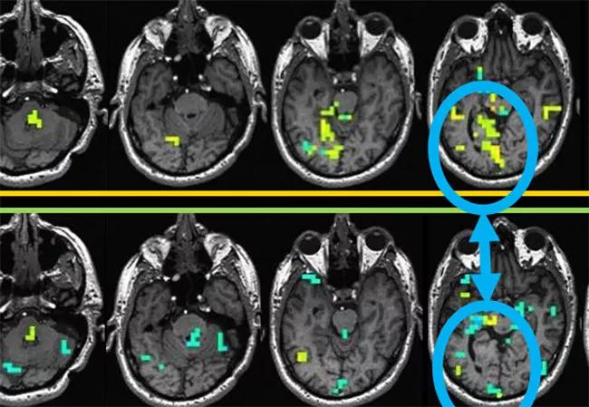
Image content: This image is available to view online.
View image online (https://assets.clevelandclinic.org/transform/baa712d9-b332-414d-9da9-0acb064bf6b4/17-NEU-4473-Jones-fMRI-650x450_jpg)
17-NEU-4473-Jones-fMRI-650×450
Advertisement
Cleveland Clinic is a non-profit academic medical center. Advertising on our site helps support our mission. We do not endorse non-Cleveland Clinic products or services. Policy
To date, the imaging techniques most applicable to neurological disease have been structural, as they can superbly delineate diseased brain regions. While MRI has been the workhorse method for the past 25 years, some diseases are often inconspicuous on structural MRI. This has spurred considerable interest in more recent MRI techniques developed to examine brain function, rather than structure, which in turn is closely related to imaging brain connectivity.
Close collaboration between Cleveland Clinic’s Imaging Institute and Neurological Institute has enabled demonstration of a new technique to image brain connectivity featuring simultaneous deep brain stimulation (DBS) electrode stimulation and functional MRI (fMRI) imaging performed at 1.5T in patients with movement disorders under general anesthesia. Robust blood-oxygen-level-dependent (BOLD) activation can be easily elicited at voltages greater than 4V. The patterns of BOLD activation include both proximal and distal brain regions, with high spatial sensitivity, and these patterns also reflect clinical efficacy.
The technique of simultaneous electrical stimulation of the brain and BOLD fMRI is generally applicable to any neurological disease that can be treated with electrical stimulation. To date, this has included epilepsy, major depression, obsessive-compulsive disorder, essential tremor and urinary incontinence. For these experiments, patients were scanned in a 1.5T intraoperative MRI scanner during DBS electrode implantation under general anesthesia. After anatomically guided implantation, a BOLD-sensitive echo planar imaging sequence was acquired during stimulation of the electrodes in a block design of 30 seconds on/off. Stimulation parameters were 2, 5 and 8V across bipolar contacts at 130 Hz. Up to four DBS-fMRI sequences were obtained during the imaging session, each with variations of stimulation parameters such as voltages, contacts and duration.
Advertisement
To date, seven patients have been studied and there have been no adverse reactions. Various stimulation parameters were explored, generally showing robust BOLD activation with voltages greater than 5V. BOLD activation included brain regions both proximal and distal to the electrodes, with patterns reflecting motor circuits, and the patterns were very sensitive to lead location. The figure presents example findings.
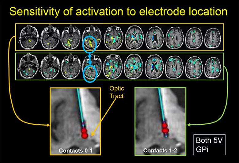
Image content: This image is available to view online.
View image online (https://assets.clevelandclinic.org/transform/dd8680ac-1aa3-4832-99a0-e5bf5998eab4/17-NEU-4473-Jones-fMRI-inset_jpg)
Figure. The top row of images shows BOLD activation maps resulting from stimulation of electrode contacts in the globus pallidus interna (GPi) regions. This stimulation, depicted in the large image on the lower left, is specifically a bipolar stimulation between contacts 0 and 1, where the red spheres in the lower left image indicate predicted regions depolarized by electrical current. Yellow-green shading indicates regions with increased BOLD activation with electrical stimulation, whereas blue regions show decreased activation with electrical stimulation. The second row of brain maps shows the results when the location of the stimulation electrodes is slightly changed, now in the adjacent contact pair 1-2. While the two rows of brain maps show activation in many brain regions, there are some notable differences, such as the right-sided visual areas of the occipital lobes, as indicated within the blue circles (fourth images in each row). This is likely due to proximity of the activation at contacts 0-1 to the optic tracts, as indicated by the arrow.
This technique of simultaneous DBS electrode stimulation and fMRI offers a possible alternative for patients desiring DBS implantation while under general anesthesia as well as functional location of electrodes to maximize clinical response and minimize side effects. This technique could easily be generalized to any functional localization electrodes during implantation.
Advertisement
Dr. Jones is Vice Chair for Research and Academic Affairs in Cleveland Clinic’s Imaging Institute and holds multiple appointments in the Neurological Institute. He acknowledges Hyun-Joo Park, PhD, and Ken Baker, PhD, of Cleveland Clinic’s Center for Neurological Restoration, as well as Pallab Bhattacharyya, PhD, and Mark Lowe PhD, of the Imaging Institute, for contributions to this report.
Advertisement
Advertisement

New study advances understanding of patient-defined goals

Testing options and therapies are expanding for this poorly understood sleep disorder

Real-world claims data and tissue culture studies set the stage for randomized clinical testing

Digital subtraction angiography remains central to assessment of ‘benign’ PMSAH

Cleveland Clinic neuromuscular specialist shares insights on AI in his field and beyond

Findings challenge dogma that microglia are exclusively destructive regardless of location in brain

Neurology is especially well positioned for opportunities to enhance clinical care and medical training

New review distills insights from studies over the past decade