Locations:

Surgical correction appropriate for subset of patients
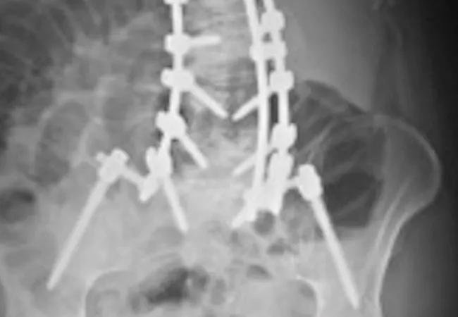
Image content: This image is available to view online.
View image online (https://assets.clevelandclinic.org/transform/b433a0ea-4bb3-4a17-a6e3-428a76eade14/16-ORT-1787-Savage-Hero-Image-650x450pxl_jpg)
16-ort-1787-savage-hero-image-650x450pxl
Advertisement
Cleveland Clinic is a non-profit academic medical center. Advertising on our site helps support our mission. We do not endorse non-Cleveland Clinic products or services. Policy
A 65-year-old female presented to Cleveland Clinic’s Center for Spine Health complaining of severe back and leg pain. She was no longer able to stand or walk for more than five to 10 minutes, and the symptoms were significantly impacting her quality of life. She went through an extended course (six months) of nonoperative care, including physical therapy, anti-inflammatory medication and epidural steroid injections. Her symptoms were refractory to these modalities, and she presented for a surgical consultation. Standing full-length radiographs revealed a severe adult degenerative scoliosis (DS) with sagittal plane imbalance (Figures 1A, B). MRI revealed severe spinal stenosis with nerve root compression throughout the curve in her lumbar spine.
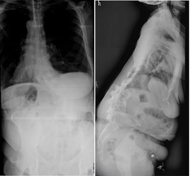
Image content: This image is available to view online.
View image online (https://assets.clevelandclinic.org/transform/603ec67f-5466-4060-a60e-491cfde040a0/16-ORT-1787-Savage-Inset-Image-01-650pxl-width_jpg)
Figures 1A, B: Preoperative radiographs reveal a profound adult DS with significant sagittal plane imbalance.
Degenerative scoliosis is a relatively common problem that often leads to significant pain and disability. An estimated 30 percent of the population has DS, defined as a curve greater than 10 degrees. This specific spinal deformity typically affects people over the age of 50 and is likely to become more prevalent as the population ages.
Not all patients with DS have clinically significant symptoms; however, a subset of patients have back pain with or without neurologic symptoms like leg pain, numbness and tingling, and even weakness. Recent studies have shown that patients with DS associated with sagittal plane imbalance (poor posture with the inability to stand upright) have significant pain and disability, and that surgical correction leads to improved health related quality of life outcome measures.
Advertisement
We performed this six-hour procedure using a posterior approach. The spine was exposed from T4 to the sacrum. We then performed a decompression from L1 to L5 to address her spinal stenosis. Posterior column osteotomies (complete facet resections) at L1-L2, L2-L3, L3-L4 and L4-L5 helped mobilize the arthritic spine. Pedicle screw instrumentation was placed from T4 to the sacrum, and we used iliac fixation to strengthen the construct. Various maneuvers were used to correct the severe deformity, and then cobalt chrome rods were placed to hold the correction (Figures 2A, B).
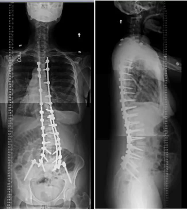
Image content: This image is available to view online.
View image online (https://assets.clevelandclinic.org/transform/eddbeff7-336b-4471-9481-8c1c2ff7329a/16-ORT-1787-Savage-Inset-Image-02-Pairing-650pxl-width_jpg)
Figures 2A and B: Postoperative radiographs reveal correction of the deformity with restoration of normal alignment.
The patient did very well postoperatively and has dramatic improvement in her posture. Her leg pain resolved immediately, and her postoperative back pain improved over six to 12 weeks. She has returned to work and is very happy with her outcome.
DS is a relatively common problem, and is often associated with significant pain and disability. Operative treatment of this complex problem can relieve symptoms and improve quality of life, but it carries significant risk and morbidity. Cleveland Clinic uses a multispecialty approach, collaborating with experts in internal medicine, bone health, anesthesia, orthopaedic surgery and neurosurgery to minimize the risk of complications and maximize patient outcomes.
Dr. Savage holds joint appointments in the Center for Spine Health in the Neurological Institute and the Department of Orthopaedic Surgery.
Advertisement
Advertisement
Advertisement

Biologic approaches, growing implants and more
Study reports zero infections in nearly 300 patients
How to diagnose and treat crystalline arthropathy after knee replacement
Study finds that fracture and infection are rare
Center will coordinate, interpret and archive imaging data for all multicenter trials conducted by the foundation’s Osteoarthritis Clinical Trial Network

Reduced narcotic use is the latest on the list of robotic surgery advantages
Cleveland Clinic specialists offer annual refresher on upper extremity fundamentals
Cleveland Clinic orthopaedic surgeons share their best tips, most challenging cases and biggest misperceptions