Locations:

High-field-strength MRI makes a difference in management
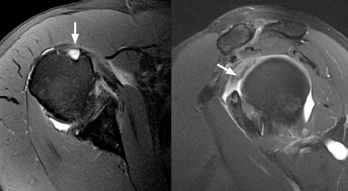
Image content: This image is available to view online.
View image online (https://assets.clevelandclinic.org/transform/391d9fbd-56de-4fd6-a4d1-ebec5e46d1b4/14-ORT-983-Polster-Hero-Image-690x380pxl_jpg)
14-ORT-983-Polster-Hero-Image-690x380pxl
By Hassana Barazi, MD, and Joshua Polster, MD
Advertisement
Cleveland Clinic is a non-profit academic medical center. Advertising on our site helps support our mission. We do not endorse non-Cleveland Clinic products or services. Policy
A 59-year-old man presented with shoulder pain of two weeks’ duration. Clinical findings were suggestive of rotator cuff pathology.
A routine noncontrast MRI examination was performed on a 3-tesla MRI machine (Siemens Verio®) using multichannel surface coils over the affected shoulder. The images demonstrated absence of the long head biceps tendon at the level of the bicipital groove without evidence of tendon dislocation, findings compatible with a complete tear with distal retraction (Figure 1). A sizable intra-articular remnant of the long head of the biceps tendon was still present and attached to the supraglenoid tubercle (Figure 2).
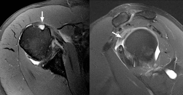
Image content: This image is available to view online.
View image online (https://assets.clevelandclinic.org/transform/e7de126d-e9e1-45ee-8109-ff63951a8cba/14-ORT-983-Polster-Inset-Image-590pxl-width_jpg)
Figure 1 (left). MRI showing absence of the long head biceps tendon at the level of the bicipital groove with no evidence of tendon dislocation, which suggests a complete tear of the tendon with distal retraction. Figure 2 (right). MRI showing absence of the long head biceps tendon at the level of the bicipital groove with no evidence of tendon dislocation, which suggests a complete tear of the tendon with distal retraction.
The management of biceps tears is generally conservative in the absence of an intra-articular remnant. Surgical management may be required when an intra-articular fragment is present. For this reason, high-quality assessment of the intra-articular biceps is important diagnostically, as it can effectively alter the course of treatment. In this case, the patient was managed conservatively with NSAIDs and physical therapy and continues to report a gradual decrease in shoulder pain with time.
Advertisement
The 3-tesla MRI system has a magnetic field strength two to three times stronger than that of standard high-field systems, allowing acquisition of images at higher speeds and with much greater signal-to-noise ratios. In combination with multichannel coils, newer software and new imaging sequences, this allows for images with significantly higher resolution of small structures. This higher resolution enables more thorough evaluation of intra-articular structures. The result is greater diagnostic certainty and ultimately better patient care.
At the time this was written, Dr. Barazi was a musculoskeletal radiology fellow in Cleveland Clinic’s Imaging Institute.
Dr. Polster is a musculoskeletal radiologist in the Department of Diagnostic Radiology with a specialty interest in sports medicine imaging.
Advertisement
Advertisement

Biologic approaches, growing implants and more
Study reports zero infections in nearly 300 patients
How to diagnose and treat crystalline arthropathy after knee replacement
Study finds that fracture and infection are rare
Center will coordinate, interpret and archive imaging data for all multicenter trials conducted by the foundation’s Osteoarthritis Clinical Trial Network

Reduced narcotic use is the latest on the list of robotic surgery advantages
Cleveland Clinic specialists offer annual refresher on upper extremity fundamentals
Cleveland Clinic orthopaedic surgeons share their best tips, most challenging cases and biggest misperceptions