Locations:

Functional assessment of 3-D-printed valve found to be feasible
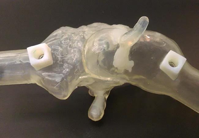
Image content: This image is available to view online.
View image online (https://assets.clevelandclinic.org/transform/83dc8e9a-02e8-4af1-aa2b-a3fdf6a0f064/Fig-1C_650x450_jpg)
Fig 1C_650x450
Three-dimensional (3-D) printing has already been used in complex congenital heart disease and in pre-procedural planning of cardiac interventions and heart surgeries. Now an innovative research project at Cleveland Clinic is revealing how the technology may help cardiologists better understand valvular pathophysiology.
Advertisement
Cleveland Clinic is a non-profit academic medical center. Advertising on our site helps support our mission. We do not endorse non-Cleveland Clinic products or services. Policy
Using 3-D printing, advanced cardiac imaging fellow Serge Harb, MD, and several Cleveland Clinic colleagues developed a functional model of a severely stenotic aortic valve. They built a circuit around the valve and successfully replicated the pressure gradients obtained through echocardiography. Manipulating the parameters allowed them to see how the valve would behave under various hemodynamic conditions.
The proof-of-concept study, presented by Dr. Harb on June 12 at the American Society of Echocardiography’s annual scientific sessions in Seattle, is among the first to include a functional assessment of a 3-D-printed valve.
“This technology holds a lot of promise,” says Dr. Harb. “We expect it will help us better understand the pathophysiology of atypical forms of aortic stenosis.”
Aortic stenosis is a relatively common disease with complex pathophysiology. The typical form, “high-flow, high-gradient,” is easily diagnosed with echo and treated via surgical valve replacement or transcatheter aortic valve replacement (TAVR). But in some forms of aortic stenosis in which the flow and/or gradient do not match the apparent severity of the valve narrowing, the underlying pathophysiology is not well understood. As a result, the optimal management approach is sometimes uncertain.
Enter 3-D printing. “We cannot adjust the heart rate and flow in a human, but we can in a 3-D-printed valve,” explains Dr. Harb. “By changing the hemodynamic settings, such as increasing the flow, we can see how the valve will behave.”
Advertisement
His team’s first step was to develop a model to test whether the pressure gradients obtained by echocardiography — the gold standard for diagnosing aortic stenosis — could be replicated by a 3-D-printed version of a stenotic aortic valve.
The aortic valve of an 83-year-old man with severe calcific aortic stenosis was chosen as the model. A 3-D reconstruction of his valve was created from high-resolution, contrast-enhanced cardiac CT images. Different materials were used for the valve leaflets and calcifications, allowing them to be easily distinguished.
To allow assessment of valve hemodynamics, the 3-D-printed valve was connected to a flow circuit containing a pulsatile pump and both pressurized and unpressurized tanks. Flow was measured using a transonic flow probe and meter. Pressures proximal and distal to the aortic model were obtained via fluid-filled sensors and signal amplifiers.
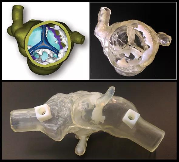
Image content: This image is available to view online.
View image online (https://assets.clevelandclinic.org/transform/a5cea0eb-e890-4981-922d-ac15bc42c39c/3-images_550x534_jpg)
Figure. Key steps in the preparation of the 3-D-printed valve model. Top left: An initial digital model based on CT scans. Top right: A 3-D-printed sample part showing leaflet and calcium detail. Bottom: Full 3-D-printed model with tubing connectors and pressure ports.
Flow, pressures and pump rate were adjusted to replicate the patient’s hemodynamic parameters at the time of his echocardiogram. The team then measured pressure gradients across the 3-D-printed valve and were able to replicate the gradients obtained by echo. The circuit settings were subsequently modified to assess how the pressure gradients changed based on varying hemodynamic conditions.
Advertisement
“We were able to measure gradients across the valve under different flow conditions and study how the valve behaves,” notes Dr. Harb.
Faced with the diagnostic challenges posed by patients with atypical forms of aortic stenosis, cardiologists require better understanding of the underlying mechanisms in order to optimize management. In this respect, 3-D printing offers tremendous promise.
“This is an exciting new field,” says cardiac imaging specialist L. Leonardo Rodriguez, MD, who served as a staff advisor on the project. “We hope that by creating a 3-D-printed model that simulates these conditions, we will be able to refine the diagnostic criteria.”
Next, the researchers will model valves from other patients to evaluate whether the process consistently replicates echo measurements. They will also seek to determine how the valve behaves when pump rate and flow are varied. “This should reveal how atypical cases of aortic stenosis with low gradient and/or low flow respond when we take control of flow,” Dr. Harb explains.
Hopefully, answers will not be long in coming. He looks forward to using the information to improve patient care.
“Printing the particular valve of an affected patient has the potential to provide personalized management,” he says, “and could be especially helpful when planning surgery or TAVR procedures.”
Advertisement
Advertisement

A sampling of outcome and volume data from our Heart & Vascular Institute

Concomitant AF ablation and LAA occlusion strongly endorsed during elective heart surgery

Large retrospective study supports its addition to BAV repair toolbox at expert centers

Young age, solid tumor, high uptake on PET and KRAS mutation signal risk, suggest need for lobectomy

Surprise findings argue for caution about testosterone use in men at risk for fracture

Residual AR related to severe preoperative AR increases risk of progression, need for reoperation

Findings support emphasis on markers of frailty related to, but not dependent on, age

Provides option for patients previously deemed anatomically unsuitable