A novel endovascular navigation technology in action
A novel endovascular navigation technology developed by Cleveland Clinic researchers has successfully completed a second round of preclinical testing on its way toward hoped-for market entry in the second half of 2017. We previewed the technology — known as the Intraoperative Positioning System (IOPS) — earlier this year, but the time seems right for a closer look in the slideshow below. Details on on the latest IOPS testing follow at the bottom of this post.
Advertisement
Cleveland Clinic is a non-profit academic medical center. Advertising on our site helps support our mission. We do not endorse non-Cleveland Clinic products or services. Policy


Advertisement


Slide 1/6
IOPS extracts the centerlines of the aorta and branch vessels from a patient’s CT images to mathematically construct a high-quality 3-D model of the relevant vasculature. Electromagnetic tracking enables use of the model in a GPS-like manner to guide navigation during minimally invasive endovascular repairs of the aorta, reducing reliance on radiation-inducing fluoroscopy.
The latest round of IOPS testing involved two more preclinical studies, in July and August, on the heels of the first study in May 2016.
“This second round of work verified our ability to navigate the aorta and its branches, selecting the celiac artery, superior mesenteric artery and renal arteries, with the use of fluoroscopy limited to verification of catheter location,” says Cleveland Clinic vascular surgeon Matthew Eagleton, MD, who served as primary investigator.
“This technology will limit the need for extensive fluoroscopy units and provide more detailed anatomy that can be imaged while navigating through it,” adds Dr. Eagleton. “It has the potential to revolutionize vascular surgery.”
Advertisement
Dr. Eagleton has a financial interest in the company developing IOPS, Centerline Biomedical, as chair of its scientific advisory board. Centerline, a Cleveland Clinic Innovations spinoff company, plans to submit IOPS for regulatory approval by the FDA, with a target U.S. market entry in the third quarter of 2017.
Advertisement
Advertisement

A sampling of outcome and volume data from our Heart & Vascular Institute
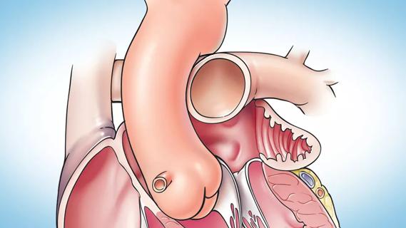
Concomitant AF ablation and LAA occlusion strongly endorsed during elective heart surgery
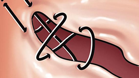
Large retrospective study supports its addition to BAV repair toolbox at expert centers
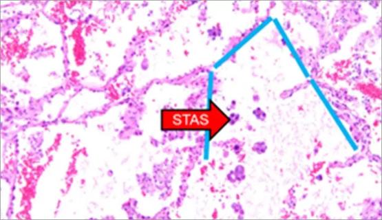
Young age, solid tumor, high uptake on PET and KRAS mutation signal risk, suggest need for lobectomy
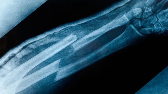
Surprise findings argue for caution about testosterone use in men at risk for fracture
Residual AR related to severe preoperative AR increases risk of progression, need for reoperation

Findings support emphasis on markers of frailty related to, but not dependent on, age
Provides option for patients previously deemed anatomically unsuitable